QUESTIONS ABOUT TREATMENT OF THYROTOXICOSIS
Loading…
Mnemonic- complications related to chronic liver disease, you can use: “H-E-A-P-S C-H-A-R-T” H – Hepatic encephalopathy E – Esophageal varices A – Ascites P – Portal hypertension S – Spontaneous bacterial peritonitis (SBP) C – Coagulopathy (bleeding disorders) H– Hepatocellular carcinoma A– Anemia R – Renal failure (hepatorenal syndrome) T – Thrombocytopenia
Cerebellar signs Mnemonic is DANISH: Dysdiadochokinesia or dysmetria Ataxia Nystagmus Intention tremor Speech (slurred or scanning) Hypotonia
GRAVE HIM Graves’ disease Radiation (iodine-induced) Adenoma (toxic adenoma) Viral thyroiditis (subacute thyroiditis) Excess iodine (Jod-Basedow phenomenon) HCG (trophoblastic disease) Iodine-containing medications (amiodarone) Multinodular goiter (toxic multinodular goiter)
THYROID’S Thyroiditis (Hashimoto’s, subacute) Hypothalamic or pituitary dysfunction Young (congenital hypothyroidism) Radiation (radiation therapy or radioactive iodine) Overdose of anti-thyroid medications Iodine deficiency/excess Drugs (lithium, amiodarone) Subacute thyroiditis
Mnemonic For Causes of CKD DIABETES (focuses on major causes): D -Diabetes (mellitus) I – Interstitial nephritis A – Autosomal dominant polycystic kidney disease (ADPKD) B – Blood pressure (Hypertension) E – Exposure to toxins (Nephrotoxins) T – Tubulointerstitial diseases E– Excessive protein intake S -Streptococcal infections (leading to glomerulonephritis)
Two CHIMPANZEES: This one is a bit longer but quite comprehensive. T- Thyroid (Hyper) C – Calcium supplementation H – Hyperparathyroidism I – Iatrogenic (drugs like thiazides or lithium, or immobility) M – Milk-alkali syndrome P – Paget’s disease of bone A – Addison’s Disease (adrenal insufficiency) N – Neoplasia (cancers) Z – Zollinger-Ellison syndrome…
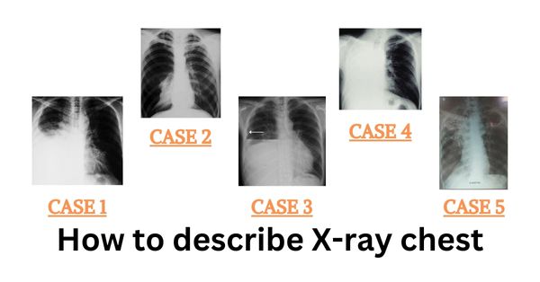
Case 1 A. Xray Chest PA view was taken in erect posture in inspiration with Adequate exposure B. Bone & Soft tissues are normal, C. Cardiac normal in position, left border is normal whereas as right side border is obscured by homogenous opacity (silhouette sign) D – Diaphram – Left diaphragm in normal position whereas…
Interpreting X-ray Chest PA view in a short time can be confusing and there lies a strong possibility of missing important findings while interpreting it. This may lead to losing numbers in table viva. Therefore below is a simple approach – how to interpret the x-ray chest PA view. …