Case 1
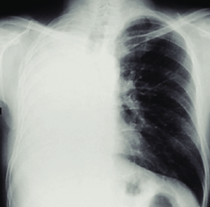
A. Xray Chest PA view was taken in erect posture in inspiration with Adequate exposure
B. Bone & Soft tissues are normal,
C. Cardiac normal in position, left border is normal whereas as right side border is obscured by homogenous opacity (silhouette sign)
D – Diaphram – Left diaphragm in normal position whereas the right side is obscured by a homogeneous opacity (silhouette sign)
E – Effusions/ extrathoracic – A homogenous opacification is noted in the right lower zone with the opacity seen to track along the lateral chest wall. The right costophrenic angle is obliterated with a meniscus noted.
F – Field, fissures, and foreign bodies – visible Lung field is normal
G – Great Vessels/ Gastric bubbles are normal
H- Hila & mediastinum – are normal
I – Interpretation – It is a skiagram of a male patient taken in an erect position PA view in the inspiration phase with normal bone, soft tissue, and cardiac position. left side of the cardiac border and left diaphragm is normal whereas a large homogenous opacity is noted in the right lower zone with the opacity seen to track along the lateral chest wall. The right costophrenic angle is obliterated with a meniscus noted on the upper border of opacity (curve formed by the upper layer of fluid).
It is a case of right-side moderate Pleural effusion
Case 2
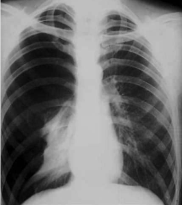
A. X-ray chest PA view was taken in erect posture in inspiration with Adequate exposure with slight rotation to the left side.
B. Bone & Soft tissues are normal.
C. Cardiac normal in position, slightly tubular with normal right & left borders.
D. Left side diaphragm is normal in position whereas the dome of the right side diaphragm is flattened and shifted downwards.
E – a large hyper lucent area is seen n right side of chest with the absence of vascular markings.
F- a thin sharp white line of visceral pleura is seen outlining the compressed right lung.
G – Normal
H – Normal
I – It is a skiagram of a male patient taken in an erect posture PA view in the inspiration phase with normal bone, soft tissue, and slight tubular cardiac shadow. A large hyperlucent area suggestive of air is seen on the right side pushing the right diaphragm down while compressing the right side lung. Mediastinum is normal in position.
It is a case of Large pneumothorax with compression collapse of the right lung.
click here to watch the video explanation of Case 2
Case 3

A. Xray Chest PA view was taken in erect posture in inspiration with Adequate exposure
B. Bone & Soft tissues are normal,
C. Cardiac normal in position, left border is normal whereas as right side border is obscured by homogenous opacity (silhouette sign)
D – Diaphram – Left diaphragm in normal position whereas the right side is obscured by a homogeneous opacity (silhouette sign)
E – Effusions/ extrathoracic – on the right side, a large homogenous opacity outlined with a sharp horizontal upper border and a hyperlucent area on the periphery of the lung field with no vascular markings and a sharp thin white line of the visceral pleural edge is seen suggesting air-fluid level.
F – Field, fissures, and foreign bodies – partial compression collapse of the right lung is seen
G – Great Vessels/ Gastric bubbles are normal
H- Hila & mediastinum – are normal
I – Interpretation – It is a skiagram of a male patient taken in an erect posture PA view in the inspiration phase with normal bone, soft tissue, and cardiac position. Left cardiac border and left diaphragm are normal whereas a large homogenous opacity is silhouetting the right border of the cardiac as well as the right diaphragm. An air-fluid level is seen on the right side of the lung suggesting a case of Right side hydro-pneumothorax with right lung collapse (partial compression collapse).
click here to watch the video explanation of Case 3
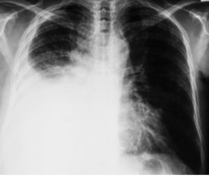
Case 4
A. X-ray chest PA view was taken in erect posture in inspiration with Adequate exposure with slight rotation to the Right side.
B. Bone & Soft tissues are normal.
C- Cardiac shadow is seen shifted to the right side with a normal left border whereas as right side border is obscured by homogenous opacity (silhouette sign).
D – Diaphram – Left diaphragm in normal position whereas the right side is obscured by a homogeneous opacity (silhouette sign).
E- The right side costodiaphragmatic angle is obliterated whereas the left side angle seen is normal.
F- A large Homogenous opacity is seen in the entire right side of the Lung with no air bronchogram whereas the left lung field is normal
G- Aortic Knuckle is seen.
H- Trachea, Hila, And Mediastinum are shifted to the right side of the lung. left Perihilar lymph node is present.
I- It is a skiagram of a male patient taken in an erect position PA view in the inspiration phase with normal bone, and soft tissue. cardiac shadow, trachea, hila, and mediastinum are shifted towards the right side of the chest where a large homogeneous opacity without air bronchogram in the entire lung field is seen. This homogenous opacity is silhouetting the right border of the cardiac as well as the right diaphragm.
It is a case of the right-sided collapse of the lung probably due to the bronchial or intrabronchial lesion with complete blockage of the major right-side bronchus.
click here to watch the video explanation of Case 4
Case 5
 A. X-ray chest PA view was taken in erect posture in inspiration with Adequate exposure with slight rotation to the Right side.
B. Bone & Soft tissues are normal,
C. Cardiac shadow is slightly tubular with mild shifting towards the right side with normal right & left borders.
D. Left side diaphragm is normal in position whereas the dome of the right side diaphragm is flattened with the medial end pulled up and merged with the shadow of the right side of the cardiac border.
E- Costodiaphragmatic angles are clear on both sides.
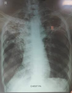
F – a large heterogeneous opacity is seen right upper lobe extending from the hila and medial part of the lung up to the periphery of the lung. A similar heterogeneous opacity is seen in the left side of the upper lung field although lesser in area but extending from the medial to periphery of the lung field. Multiple small thick-walled cavitary lesions with irregular margins are seen on both the upper lung lobes amidst the heterogeneous opacity.
G. Aortic Knukle is seeen normal
H – Trachea, hila, and mediastinum are shifted towards the right side.
I – It is a skiagram of a male patient taken in an erect position PA view in the inspiration phase with normal bone, and soft tissue. cardiac shadow, trachea, hila, and mediastinum are shifted towards the right side of the chest whereas the medial part of right side diaphragm is pulled upwards. The heterogenious opacity and multiple thick walled irregularly outlined cavitatory lesions on seen both the lung fields (right > left).
It is case of b/l upper lobe Fibrocavitatory disease with fibrosis more on right side as compared to left side of lung – cause? Pulmonary tuberculosis
click here to watch the video explanation of Case 5